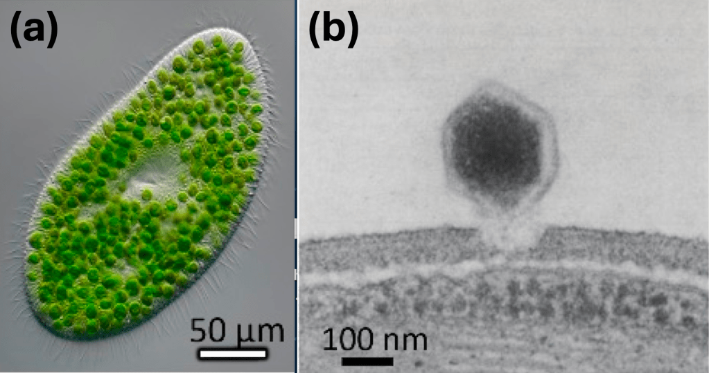
All organisms on Earth experience infection by viruses. This includes the estimated one trillion microbial species that inhabit every part of Earth’s surface, from deep-sea hydrothermal vents to the clouds (Locey and Lennon 2016). If we assume that each of these one trillion microorganisms hosts at least one virus, we begin to appreciate the vast diversity of the global virosphere. Unfortunately (or perhaps excitingly), much of this diversity remains undescribed, with only about 17,500 viral species recognized by the International Committee on Taxonomy of Viruses (ICTV; https://ictv.global/msl).
Though viruses infecting bacteria have long been the focus of virologists, by the late 20th century, it became clear that viruses were also infecting another group of microorganisms: the eukaryotic microalgae (Rodrigues 2026). In the 1970s, Kawakami and colleagues observed microscopic particles, later found to be viruses, contained inside a ciliate called Paramecium bursaria (Kawakami and Kawakami 1978). But these viruses were not infecting P. bursaria. In fact, they were infecting a microalga in the family Chlorellaceae, which lives symbiotically within P. bursaria and provides the ciliate with nutrients via photosynthesis. These algae are commonly referred to as zoochlorellae and exist as symbionts in protozoans and metazoans. As scientists are apt to do, they promptly sought to classify this new virus, placing it in the novel genus Chlorovirus.
Since this discovery, chloroviruses have become among the most well-studied algal viruses and have revealed an unprecedented level of complexity in the ecology of aquatic viruses. Chloroviruses are currently classified within the viral family Phycodnaviridae, which is found in the order Algavirales (though this may change as viral taxonomy is an ever-evolving field). Chloroviruses are highly diverse, with three primary groups having been described thus far. Their names were initially chosen to correspond to the Chlorella strain that they could infect (Jeanniard et al. 2013). The first is the NC64A viruses, which infect C. variabilis strain NC64A. More recently, chloroviruses infecting C. variabilis strain Syngen 2-3 were isolated and designated OSy-viruses, for “Only Syngen” (Quispe et al. 2017). Following the same nomenclature, SAG chloroviruses infect the C. heliozoae strain SAG3.83 (Fitzgerald, Graves, Li, Hartigan, et al. 2007), and Pbi viruses were isolated from Micractinium conductrix strain Pbi (Fitzgerald, Graves, Li, Feldblyum, et al. 2007). Though this classification system seemed straightforward at first, it began to break down as more viruses were discovered. For instance, there is now evidence that OSy-viruses infect more than just the Syngen 2-3 Chlorella strain, meaning that the name “Only Syngen virus” is inaccurate (Carvalho et al. 2024). Hence, scientists recently proposed designating three subgenera, classifying chloroviruses into Alphachloroviruses (NC64A Virus and OSy viruses), Betachloroviruses (Pbi viruses), and Gammachloroviruses (SAG viruses; Carvalho et al. 2024).
Of the 6 other genera grouped under Phycodnaviridae, the greatest number of chlorovirus sequences are available in the open-access database at the National Center for Biotechnology Information (NCBI) GenBank. Importantly, many chloroviruses have been successfully isolated from numerous inland aquatic environments across North America, Europe, and Asia (Jeanniard et al. 2013). Having viral isolates provides significantly more information than simply relying on viral sequences. Namely, researchers can use these isolates in controlled experiments to test specific hypotheses. Because of this, chloroviruses have helped to reveal complicated aspects of viral ecology.
In this post, I will highlight a few particularly interesting ecological interactions. Note that much of this research was conducted using the chlorovirus PBCV-1 (Chlorovirus vanettense) infecting endosymbiotic chlorella found in P. bursaria.
Work by Dunigan and colleagues showed that chloroviruses attract P. bursaria, presumably by releasing an unknown infochemical (Dunigan et al. 2019). This may be because chloroviruses can attach to the surface of P. bursaria, essentially hitching a ride on the ciliate. This may increase the virus’s probability of encountering its host, which lives inside of P. bursaria (DeLong, Van Etten, and Dunigan 2023). For this encounter to occur, however, the ciliate must rupture, exposing the zoochlorellae residing inside of it. This happens when other members of the microbial community feed on P. bursaria (referred to as predator catalysis). Interestingly, the predator type that consumes P. bursaria may shape the dynamics of the ensuing bloom. For instance, DeLong and colleagues reported that copepods feeding on P. bursaria only partially digested their prey, preserving the endosymbionts and leading to a rapid increase in the number of viral particles (DeLong et al. 2016). A similar process occurred when ciliates were preyed upon by smaller Didnium nasutum cells where the rupture of P. bursaria cells exposed zoochlorellae to viruses. On the other hand, larger D. nasutum cells engulfed the ciliate whole, such that the chloroviruses were less easily able to access the endosymbiotic algae (DeLong et al. 2018) and leading to a smaller bloom. P. bursaria may also be attracted to chloroviruses out of a desire to consume them (virovory). Though not particularly calorically dense, viruses are contain high amounts of limiting nutrients like nitrogen and phosphorus (DeLong, Van Etten, and Dunigan 2023). Indeed, research has shown that protist growth when fed only viruses aligns with the rate of growth when fed other food sources (DeLong, Van Etten, Al-Ameeli, et al. 2023).
These findings highlight complex connections among many members of aquatic communities and raise fascinating questions. First, why do the P. bursaria cells gravitate towards chloroviruses? If the zoochlorellae are generally assumed to benefit the ciliate by providing nutrients, why would P. bursaria seek to remove them? Do the zoochlorellae respond in some way to the attachment of chloroviruses on P. bursaria cells? For the ciliate, could the benefits of consuming chloroviruses outweigh the risk of losing its endosymbionts? Are there situations in which zoochlorellae harm the ciliate? Of course, the biggest question is whether such complexity can be found in other algae-virus interactions. Given the vast number of undescribed viruses, it is highly likely that similar dynamics occur elsewhere and have yet to be uncovered.
References:
Carvalho, João Victor R. P., Roger M. Carlson, Jayadri Ghosh, et al. 2024. “Genomics and Evolutionary Analysis of Chlorella Variabilis- Infecting Viruses Demarcate Criteria for Defining Species of Giant Viruses.” Journal of Virology 98 (11): e00361-24. https://doi.org/10.1128/jvi.00361-24.
DeLong, John P., Zeina Al-Ameeli, Garry Duncan, James L. Van Etten, and David D. Dunigan. 2016. “Predators Catalyze an Increase in Chloroviruses by Foraging on the Symbiotic Hosts of Zoochlorellae.” Proceedings of the National Academy of Sciences 113 (48): 13780–84. https://doi.org/10.1073/pnas.1613843113.
DeLong, John P., Zeina Al-Ameeli, Shelby Lyon, James L. Van Etten, and David D. Dunigan. 2018. “Size-Dependent Catalysis of Chlorovirus Population Growth by A Messy Feeding Predator.” Microbial Ecology 75 (4): 847–53. https://doi.org/10.1007/s00248-017-1106-8.
DeLong, John P., James L. Van Etten, Zeina Al-Ameeli, Irina V. Agarkova, and David D. Dunigan. 2023. “The Consumption of Viruses Returns Energy to Food Chains.” Proceedings of the National Academy of Sciences 120 (1): e2215000120. https://doi.org/10.1073/pnas.2215000120.
DeLong, John P., James L. Van Etten, and David D. Dunigan. 2023. “Lessons from Chloroviruses: The Complex and Diverse Roles of Viruses in Food Webs.” Journal of Virology 97 (5): e00275-23. https://doi.org/10.1128/jvi.00275-23.
Dunigan, David D., Maitham Al-Sammak, Zeina Al-Ameeli, Irina V. Agarkova, John P. DeLong, and James L. Van Etten. 2019. “Chloroviruses Lure Hosts through Long-Distance Chemical Signaling.” Journal of Virology 93 (7): e01688-18. https://doi.org/10.1128/JVI.01688-18.
Fitzgerald, Lisa A., Michael V. Graves, Xiao Li, James Hartigan, et al. 2007. “Sequence and Annotation of the 288-Kb ATCV-1 Virus That Infects an Endosymbiotic Chlorella Strain of the Heliozoon Acanthocystis Turfacea.” Virology 362 (2): 350–61. https://doi.org/10.1016/j.virol.2006.12.028.
Fitzgerald, Lisa A., Michael V. Graves, Xiao Li, Tamara Feldblyum, James Hartigan, and James L. Van Etten. 2007. “Sequence and Annotation of the 314-Kb MT325 and the 321-Kb FR483 Viruses That Infect Chlorella Pbi.” Virology 358 (2): 459–71. https://doi.org/10.1016/j.virol.2006.08.034.
Jeanniard, Adrien, David D. Dunigan, James R. Gurnon, et al. 2013. “Towards Defining the Chloroviruses: A Genomic Journey through a Genus of Large DNA Viruses.” BMC Genomics 14 (1): 158. https://doi.org/10.1186/1471-2164-14-158.
Kawakami, Hisako, and Noboru Kawakami. 1978. “Behavior of a Virus in a Symbiotic System, Paramecium Bursaria —Zoochlorella.” The Journal of Protozoology 25 (2): 217–25. https://doi.org/10.1111/j.1550-7408.1978.tb04399.x.
Locey, Kenneth J., and Jay T. Lennon. 2016. “Scaling Laws Predict Global Microbial Diversity.” Proceedings of the National Academy of Sciences 113 (21): 5970–75. https://doi.org/10.1073/pnas.1521291113.
Quispe, Cristian F., Ahmed Esmael, Olivia Sonderman, et al. 2017. “Characterization of a New Chlorovirus Type with Permissive and Non-Permissive Features on Phylogenetically Related Algal Strains.” Virology 500 (January): 103–13. https://doi.org/10.1016/j.virol.2016.10.013.
Rodrigues, Rodrigo A. L. 2026. “Diversity and Taxonomy of Giant Algal Viruses: Chloroviruses Lead the Way.” Journal of Virology 100 (6): e01688-25. https://doi.org/10.1128/jvi.01688-25.


Leave a comment